Skip to main content
Skip to main navigation menu
Skip to site footer
Open Menu
Login
Register
SUBMIT YOUR PAPER
Petunjuk Penulisan
Biaya Penulis
Contact
QUICK LINK
Editorial Team
Reviewer
Fokus dan Ruang Lingkup
Kebijakan Plagiarisme
Kebijaksanaan Arsip
Pernyataan Akses Terbuka
POLICIES
Etika Publikasi
Peer Review Process
Kebijakan Akses Terbuka
Hak Cipta Lisensi Jurnal
About
About the Journal
Submissions
Privacy Statement
OTHER
Current
Archives
Copyright Notice
Indexsasi Jurnal
Bekerjasama dengan:
Search
Register
Login
Home
/
Login
Login
Required fields are marked with an asterisk:
*
Login
Username
*
Required
Password
*
Required
Forgot your password?
Keep me logged in
Login
Register
Menu
Accreditation
Journal Metric & Achievement
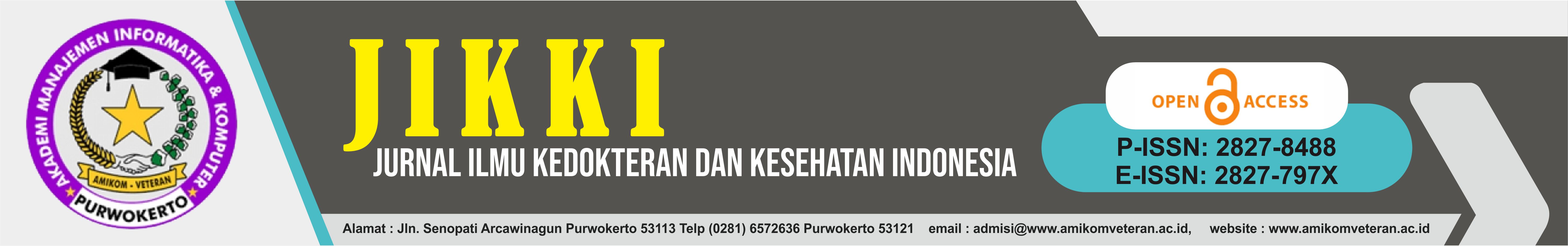
P-ISSN : 2827-8488
E-ISSN : 2827-797X
MENU
Kontak
Dewan Editorial
Reviewers
Proses Peer Review
Fokus dan Ruang Lingkup
Etika Publikasi
Kebijakan Akses Terbuka
Kebijakan Arsip
Pernyataan Akses Terbuka
Kebijakan Plagiarisme
Hak Cipta Lisensi Jurnal
Petunjuk Penulisan
Biaya Penulis
Indexing
TOOLS